
I guess quite a few of you will have had to undergo a Magnetic Resonance Imaging scan over the past few years. As you lie in the machine’s tube with headphones on to muffle the loud ‘clacking’ of the machine, have you ever wondered how it works? No? Then you can quickly go on to not reading the comments! Yes? Then please read on!
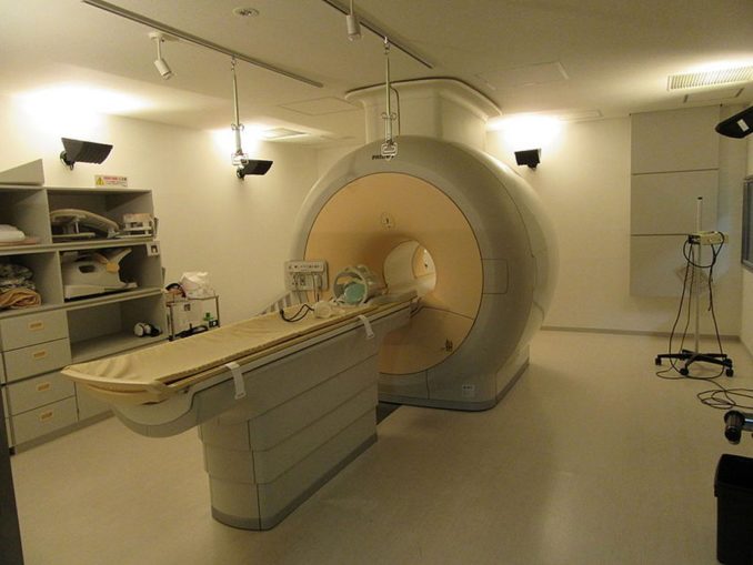
Walter Davies, CC BY-SA 4.0, via Wikimedia Commons
Magnetic Resonance, or more exactly, Nuclear Magnetic Resonance, was first demonstrated in the 1940s, leading to a Nobel prize for Israel Rabi in 1945, and was further developed by Purcell and Bloch who also won Nobel prizes in 1954. The ‘Nuclear’ bit of the name has now been dropped from MRI because it is apparently too scary a word for patients!
Here comes some science. If you put a sample of water in a strong magnetic field, the proton in the nucleus of the hydrogen atoms (the H in H2O) lines up in the field according to the quantum mechanical property of ‘spin’. Protons have a spin value of ½, so only have two possible positions, up or down in the magnetic field.
The alignment can be flipped by transmitting high-powered radio wave pulses at exactly the right frequency for the strength of the magnet to obtain a measurable signal. This is the ‘resonance’ bit of the name. Resonance is also how opera singers can break a wine glass by singing a note at exactly the right resonant frequency to make the glass vibrate and shatter.
You’ll be pleased to know that the maths of MRI is very simple:
f = gB
where f is the radio wave frequency in megahertz (MHz), B is the magnetic field strength in tesla (T), and g is the gyromagnetic ratio which is fixed for every kind of nucleus. For the protons in H, this value is 42.6 MHz/T.
You can see from the maths that a proton in a different magnetic field will have a different radio frequency. MRI therefore uses a magnetic field gradient to work. The diagram shows how you can create a one-dimensional image of three tubes of water in a field gradient as the frequency of the radio waves tells you where the tubes are positioned! This discovery led to more Nobel prizes, for Lauterbur and Mansfield, in 2003.
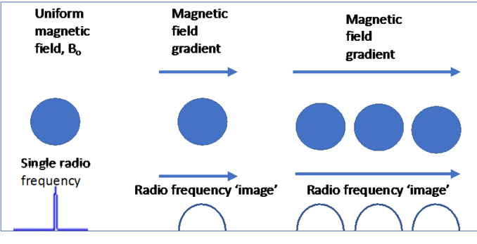
© Richard Puller 2022
So lets look more closely at the MRI scanner you’re lying in!
The tube you slide into in the radiology department is actually a very powerful magnet, made from superconducting wire cooled by liquid helium to -269 oC. More Nobel prizes behind these discoveries too! For MRI, the magnet strength is usually between 0.5T and 1.5T, that’s 10,000 to 30,000 times stronger than the Earth’s magnetic field. The MRI scanner is mainly imaging the H protons in the water in the different organs of your body, which means the radio frequencies are between 21 and 63 MHz, well away from the FM band on your radio, which is 90-110 MHz.
The magnet is why you have to take off anything metal, like watches, and get asked all those questions about whether you have any metal implants in your body. I know to my cost that magnets are very efficient at ruining watches and wiping bank cards!
To create a three-dimensional image, we need three different field gradients: up-down, left-right and along the tube. These are formed by electrical coils inside the tube. As the gradient coils switch on and off, they make the mechanical clacking noise you hear. Different scans need complex switching of the gradients and radio frequency pulses, which is why the sounds you hear seem to drift in and out of loudness and have different patterns.
There’s also a radio transmitter in the magnet tube and a radio signal detector that will be placed around your body depending where needs to be imaged. The signals are collected and processed to allow the radiographer to select ‘slices’ through your body and zoom in on the area of interest to provide the final scans to the radiologist for diagnosis.

everyone’s idle, CC BY-SA 2.0, via Wikimedia Commons
Different organs, bones, blood, etc, have different amounts of water, so show up as regions of different contrast in the image. The radio transmitter pulse sequences can also be varied to provide extra contrast due to ‘relaxation’ of the radio signals in different body tissues. Sometimes a ‘contrast agent’ made from the metal gadolinium is given to you by injection to highlight specific features that the clinicians may be looking for.
Finally, here are two MRI images of the inside of my head, before and after the 6 hour surgery to remove the large pituitary tumour that was sending me blind and would have killed me within six months. The tumour is the large mottled grey blob in the red circle. The keyhole technology would have been impossible without detailed 3-dimensional MRI scans to guide the neurosurgery team.
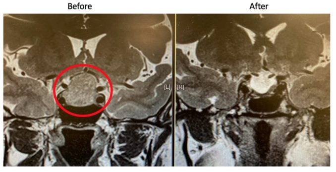
© Richard Puller 2022
Thankfully for me, and I’m sure for many of you and your loved ones, some brilliant scientists and engineers made these amazing discoveries in the last 80 years to create the MRI scanners we take for granted today.
© Richard Puller 2022
